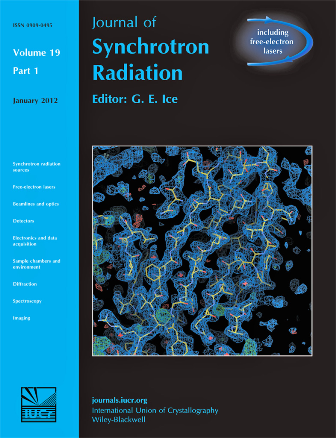
Research from the Institute of Integrative Biology has been published in the latest issue of the Journal of Synchrotron Radiation.
The Liverpool team, led by Dr Richard Strange from Structural and Chemical Biology, reported on the capabilities of current state-of-the-art software and x-ray crystallography beamlines at synchrotrons worldwide for routine automated protein structure solution using the ‘S(sulphur)-SAD phasing’ method.
X-ray protein crystallography is a powerful method for visualising in three dimensions the structures of proteins at the atomic level, enabling us to study in detail their functions, chemical reactions (eg enzyme catalysis), interactions with other types of molecules like drug-binding, and interactions with other proteins.
Protein crystallography has had a significant impact in science and society and has become a key tool for the rational design of new drug molecules and for design of catalysts for science and industry.
The ‘S-SAD’ method in protein crystallography exploits the ‘anomalous’ x-ray scattering properties of the sulphur atoms present in proteins to solve the so called ‘phase problem’. Since sulphur atoms are present in nearly every protein, as methionine or cysteine amino acids, the method is in principle applicable to any protein crystal.
Dr Strange said: “This study, which also made the front cover of the journal, shows that the vast potential this method has for solving novel protein structures is yet to be fully exploited. Improvements to existing synchrotron beamlines or new beamlines are needed to achieve greater success. “
The team are currently engaged with scientists at the Diamond Light Source in this area, with a joint PhD studentship related to Diamond’s phase III ‘long wavelength’ beamline, which is due for operation in mid 2013.
Doutch, J, Hough, M.A, Hasnain, SS and Strange, RW ‘Challenges of Sulfur SAD phasing as a routine method in macromolecular crystallography’, Journal of Synchrotron Radiation, 2012, Volume 19, pages 19-29.
