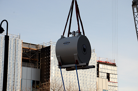
The Centre for Preclinical Imaging has taken delivery of the University of Liverpool’s first high-field pre-clinical magnetic resonance imaging (MRI) scanner.
The £1.7m, 9.4 Tesla scanner can perform very high resolution imaging, enabling the anatomy and physiology of the tissues and organs of small animals to be investigated non-invasively.
It is the latest addition to the newly developed Centre for Preclinical Imaging, joining an already state-of-the-art facility that also includes an IVIS Spectrum for bioluminescence/fluorescence; a Multi-Spectral Optoacoustic Tomography instrument and a high-frequency Ultrasound Scanner. Future plans are in place to acquire a Single Photon-Emission Computed Tomography (SPECT).
Professor Harish Poptani, Chair of Pre-Clinical Imaging in the University’s Institute of Translational Medicine, said: “The development of a Preclinical Imaging Centre will act as a catalyst for fostering collaborative research partnerships within the University, as well as with neighbouring academic institutions and industrial research programmes.”
The scanner was awarded as part of a Medical Research Council (MRC) Capital Award linked to the UK Regenerative Medicine Platform (UKRMP). The UKRMP Safety and Efficacy Hub, led from Liverpool, is developing novel strategies for evaluating the safety and efficacy of regenerative medicine therapies, and its work will be significantly enhanced by the arrival of this new equipment.
As well as being able to monitor organ structure and function in health and disease, it can also be used to track cells labelled with contrast agents such as Superparamagnetic Iron Oxide Nanoparticles (SPIONs). This, alongside numerous applications, will not only aid the further development and refinement of regenerative medicine therapies, but will also facilitate research in other areas such as developmental biology, neurobiology, physiology, pharmacology and cancer.
The Institute of Translational Medicine is currently planning a workshop to advise the research community of potential applications of these new imaging modalities.